Enlarged Human Ear, 3 Parts Ear Anatomy Model


Print Profile(1)

Description
Boost Me (for free)
Want more educational models? Boost my work, so I can quit my job!!! : )
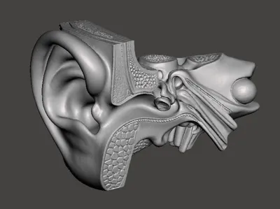
🦻 Ear Enlarged Anatomy Model – 3D Printable Educational Set
Bring human anatomy to life with this highly detailed, enlarged 3D printable model of the human ear, designed for educators, students, audiologists, and curious makers. This multipart model showcases the full auditory pathway—from the outer ear to the intricate structures of the inner ear—with labeled components for easy reference and assembly.
I modeled it after this example, and you can find all the parts here, including for the cochlear structure: https://www.amazon.com/dp/B0DK6YH11L/ref=sspa_dk_detail_0?pd_rd_i=B0DK6YH11L&pd_rd_w=SyUNU&content-id=amzn1.sym.8c2f9165-8e93-42a1-8313-73d3809141a2&pf_rd_p=8c2f9165-8e93-42a1-8313-73d3809141a2&pf_rd_r=4530ABMJ7EDWVMJSECMB&pd_rd_wg=YH1Gx&pd_rd_r=a93a490f-960d-4984-a0a4-47ab7ff9009c&sp_csd=d2lkZ2V0TmFtZT1zcF9kZXRhaWw&th=1
🔍 Features:
- Modular Design: Each section prints separately for easy painting, labeling, and classroom demonstration.
- Educational Labels Included: All anatomical components listed below are modeled with realistic proportions and surface detail.
- Optimized for FDM Printing: Supports minimal overhangs and includes flat bases for display or mounting.
📦 Includes:
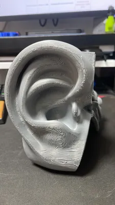
A. External Ear
- Auriculae (Pinna) – Curved outer ear structure with realistic folds
- External Acoustic Meatus – Ear canal leading to the tympanic membrane
- Periosteum – Outer bone layer modeled with subtle texture
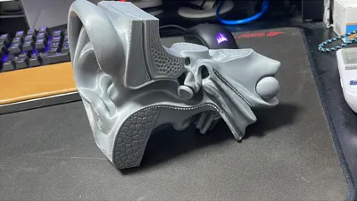
B. Middle Ear
- Tympanic Cavity – Chamber housing the ossicles
- Mastoid Antrum – Posterior cavity with honeycomb bone texture
- Eustachian Tube – Sloped channel connecting to the nasopharynx
- Ear Ossicles:
- Malleus – Hammer-shaped bone
- Incus – Anvil-shaped connector
- Stapes – Stirrup-shaped final link to the inner ear
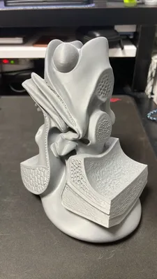
C. Internal Ear
- Cochlea – Spiral-shaped organ with visible internal chambers
- Vestibule:
- Oval Window – Entry point for sound vibrations
- Round Window – Pressure release membrane
- Semicircular Canals:
- Anterior Canal
- Posterior Canal
- Lateral Canal
- Vestibulocochlear Nerve – Modeled with branching fibers and root structure
D. Also includes a 3d printable cochlear structure
🧠 Ideal For:
- Biology and medical classrooms
- Audiology and ENT training
- Maker projects focused on anatomical modeling
- Scout STEM demos or interactive learning stations
🖨️ Print Tips:
- Recommended layer height: 0.15–0.2 mm
- Supports: Minimal (only for cochlea and canals)
- Material: PLA or PETG for best detail retention
- Assembly: Snap-fit or glue recommended for ossicles and nerve structures
License
You shall not share, sub-license, sell, rent, host, transfer, or distribute in any way the digital or 3D printed versions of this object, nor any other derivative work of this object in its digital or physical format (including - but not limited to - remixes of this object, and hosting on other digital platforms). The objects may not be used without permission in any way whatsoever in which you charge money, or collect fees.
Comment & Rating (28)